Anatomy Of Chest X Ray / Pneumoconiosis Identification In Chest X Ray Films With Cnn Based Transfer Learning Springerlink / Cxrs made easy, second edition.
Anatomy Of Chest X Ray / Pneumoconiosis Identification In Chest X Ray Films With Cnn Based Transfer Learning Springerlink / Cxrs made easy, second edition.. Xray is a type of radiography and most widely used investigation. Living anatomy of the chest for 1st year medical students original version compiled by dr. Therefore, knowing the basics and pathologies in the ed setting is very important. Major structures are shown in fig. The radiologist needs to know both the structures within the mediastinum forming the mediastinal margins and the.
L the portion of the left lung that corresponds anatomically to the right middle lobe is incorporated into the left upper lobe. Heart and great vessels — assessment of the cardiovascular anatomy includes assessment of heart and chamber size as well as the position and size of the great. Conclusion of living anatomy of the chest congratulations! Labeled chest radiographs teaching radiologic anatomy with a level of detail appropriate for medical students. The interpretation of a chest film requires the understanding of basic principles.
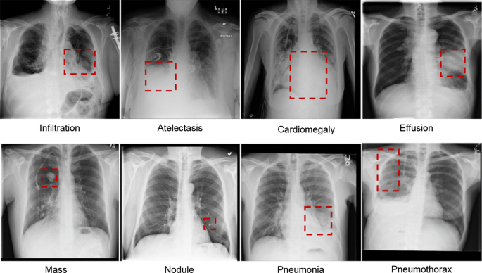
Computer Aided Detection In Chest Radiography Based On Artificial Intelligence A Survey Biomedical Engineering Online Full Text from media.springernature.com Cxrs made easy, second edition. Air spaces normally seen in. The interpretation of a chest film requires the understanding of basic principles. The radiologist needs to know both the structures within the mediastinum forming the mediastinal margins and the. Chest radiographs are the most common film taken in medicine. Living anatomy of the chest for 1st year medical students original version compiled by dr. Anatomy of the respiratory tract. It is almost always the first imaging study ordered to evaluate for pathologies of the thorax, although further diagnostic imaging, laboratory tests.
Each of these anatomical structures should be viewed using a systematic approach.
Abcde aproach the anatomy of the heart can appear artificially larger due to this image orientation. In fact every radiologist and pulmonary physician should be an expert in chest film reading. Chest radiographs are the most common film taken in medicine. Living anatomy of the chest for 1st year medical students original version compiled by dr. Christopher clarke and anthony dux. Anatomy of a chest x ray. There are also important structures that are obscured or become visible. This imaging method can also check how a patient is responding to specific treatments. A collection of anatomy notes covering the key anatomy concepts that medical students need to learn. Is there any inhaled foreign body? Anatomy of the respiratory tract. Next, a good inspiration film should show at least the 10th or 11th. Both lungs should be well expanded and similar in volume.
Xray is a type of radiography and most widely used investigation. There are also important structures that are obscured or become visible. Living anatomy of the chest for 1st year medical students original version compiled by dr. In this article we will focus on: In fact every radiologst should be an expert in chest film reading.
Cardiomediastinal Outlines On Chest X Ray Radiology Case Radiopaedia Org from prod-images-static.radiopaedia.org Gillian lieberman forthe harvard 62. Look for lung and pleural pathology. Labeled chest radiographs teaching radiologic anatomy with a level of detail appropriate for medical students. Next, a good inspiration film should show at least the 10th or 11th. L the portion of the left lung that corresponds anatomically to the right middle lobe is incorporated into the left upper lobe. Air spaces normally seen in. It first appears too complicated to read the chest xrays because we barely know what. In fact every radiologist and pulmonary physician should be an expert in chest film reading.
Therefore, knowing the basics and pathologies in the ed setting is very important.
Anatomy of the respiratory tract. You have completed this module. Is there any inhaled foreign body? The following normal chest radiographs show the normal chest anatomy. In fact every radiologst should be an expert in chest film reading. It is almost always the first imaging study ordered to evaluate for pathologies of the thorax, although further diagnostic imaging, laboratory tests. The interpretation of a chest film requires the understanding of basic principles. Each of these anatomical structures should be viewed using a systematic approach. Labeled chest radiographs teaching radiologic anatomy with a level of detail appropriate for medical students. Therefore, knowing the basics and pathologies in the ed setting is very important. Chest radiographs are the most common film taken in medicine. Xray is a type of radiography and most widely used investigation. Major structures are shown in fig.
It is almost always the first imaging study ordered to evaluate for pathologies of the thorax, although further diagnostic imaging, laboratory tests. Is there any inhaled foreign body? In this article we will focus on: Chest radiographs are the most common film taken in medicine. Heart and great vessels — assessment of the cardiovascular anatomy includes assessment of heart and chamber size as well as the position and size of the great.
The Chest X Ray A Systematic Teaching Atlas Von Matthias Hofer from images2.medimops.eu Chest radiographs are the most common film taken in medicine. The interpretation of a chest film requires the understanding of basic principles. Therefore, knowing the basics and pathologies in the ed setting is very important. Cxrs made easy, second edition. L the portion of the left lung that corresponds anatomically to the right middle lobe is incorporated into the left upper lobe. A method for examining a chest. Conclusion of living anatomy of the chest congratulations! Labeled chest radiographs teaching radiologic anatomy with a level of detail appropriate for medical students.
In fact every radiologist and pulmonary physician should be an expert in chest film reading.
A collection of anatomy notes covering the key anatomy concepts that medical students need to learn. It is almost always the first imaging study ordered to evaluate for pathologies of the thorax, although further diagnostic imaging, laboratory tests. This imaging method can also check how a patient is responding to specific treatments. Look for lung and pleural pathology. Common symptoms that can be diagnosed using chest. Gillian lieberman forthe harvard 62. Living anatomy of the chest for 1st year medical students original version compiled by dr. Is there any inhaled foreign body? Both lungs should be well expanded and similar in volume. Christopher clarke and anthony dux. In fact every radiologst should be an expert in chest film reading. Anatomy of the respiratory tract. Heart and great vessels — assessment of the cardiovascular anatomy includes assessment of heart and chamber size as well as the position and size of the great.
Christopher clarke and anthony dux anatomy of chest. Abcde aproach the anatomy of the heart can appear artificially larger due to this image orientation.


0 Comments